Antibody Identity card
Antibody information [CD1d ]
SNOW631C
- Clone name SNOW631C
- Description Rat monoclonal
- Antigen used RBL1-CD1d transfected cells and CD1d-HIS
- Epitope GTFSDQQWETLQHIF
- Isotype IgG2a
- Confirmed species reactivity Human
- Ab used CNIO purified antibody (3.4mg/ml)
| APPLICATION | Recommended concentration | Status | Protocol |
|---|---|---|---|
| Western blotting (WB) | Neat tissue culture supernatant | Working | Western Blotting (WB) |
| Immunocytochemistry (ICC) | Neat tissue culture supernatant | Working | ICC in frozen tissue and cytospin preparation |
| Immunohistochemistry (IHC-P) | 250ug/ml | Working | BOND-MAX automated immunohistochemistry |
| Immunoflourescence (IF) | 250ug/ml | Working | Immunofluorescence staining |
| Flow cytometry (FC) | 0.22 mg/ml | Working | Flow cytometry |
| IHC-P Species | Not tested |
Ab ID Card application validation / characterisation
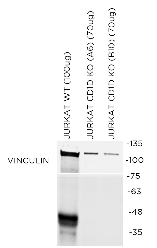
WB Validation
Over-expression/cross reactivity

Gene inactivation
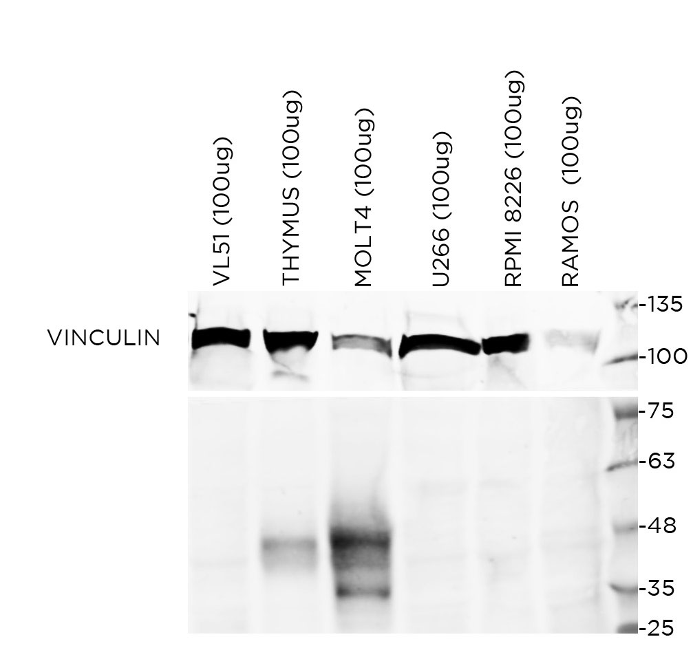
WB Characterisation
Endogenous expression
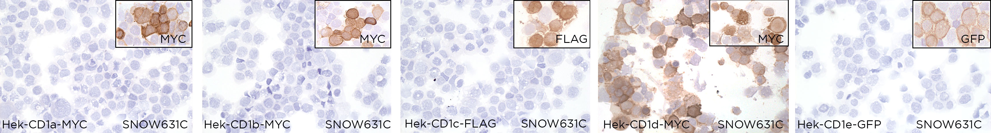
ICC / ICC-P Validation
Over-expression/cross reactivity
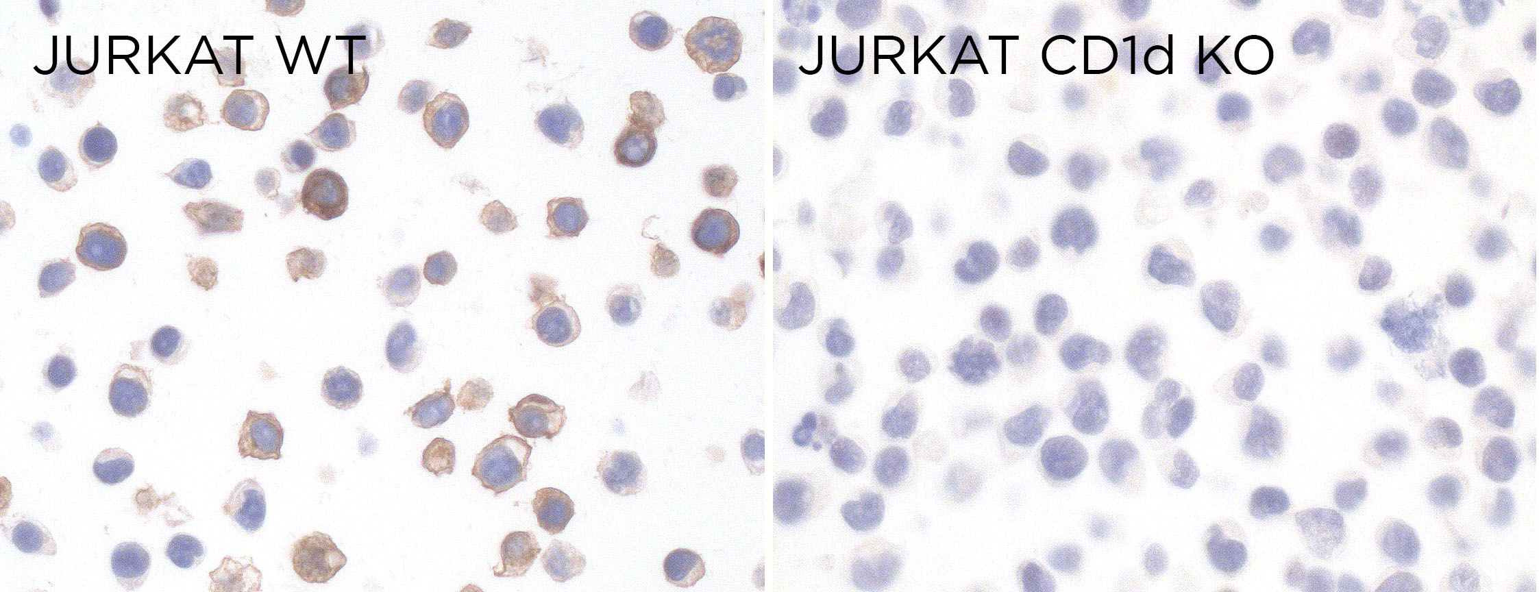
Gene inactivation
IHC-P Characterisation
Endogenous expression

IF Characterisation
Endogenous expression

FC Validation
Over-expression/cross reactivity
Not working
Gene inactivation
Not working
FC Characterisation
Endogenous expression
Not working
IHC-P Domestic species
Not tested
IHC-P Wild species
Not tested