Protocols
Hybridoma Production
A hybridoma is a cell line arising from one hybrid cell that is capable of secreting a monoclonal antibody specific to one epitope of your antigen permanently in culture. The hybrid cell is produced through the fusion of specific antibody producing B-cell from an immunized animal (usually a mouse, rat or rabbit) and which has a finite lifespan, with a cell from an “immortal” cultured myeloma cell line (e.g. mouse NS-1 or NS-0).
Production of a mouse hybrid cell
During the fusion process, B cells are isolated from the mouse spleen, mixed with the mouse myeloma cell line and fusion is induced with polyethylene glycol (PEG, see Appendix I). (The relevant myeloma line is used when B cells from other animal species are used). The resulting hybridomas are then cultured in tissue culture medium containing Hypoxathine, Aminopterin, Thymidine (HAT), a step which kills any unfused myeloma cells that might outgrow the other weaker hybridoma cells. Unfused B cells have limited powers of division and will die off naturally in culture. Ten days after the fusion process, culture supernatant is collected and tested for the presence of the desired antibody.
Schematic representation of cell fusion

Equipment needed
- A sterile environment, in which to prepare and handle cells (laminar flow or class II cabinet)
- An incubator set to 37ºC, with 5% CO2 and humidity of 95%
- An inverted microscope
- A 37ºC waterbath which can be placed in the cabinet
- A centrifuge with a swing-out rotor
- Sterile dissecting instruments -ideally two sets – each consisting of two pairs scissors and forceps (one curved and one blunt ended).
- 75 ml tissue Corning culture flasks – Ref. 15430641
- 24 well Falcon plates - Ref. 353047
- Sterile pipettes
- Pipette filler
- Sterile pasteur pipettes
- A timer
Medium and other reagents (See Appendix A for more details)
- RPMI 1640 bicarbonate buffered, with L-Glutamine (Lonza Ref. BE12-702F)
- RPMI 1640 Hepes buffered, without L-Glutamine
- Good quality (batch tested) Foetal Bovine Serum (Genycell Ref. GCS0101-500)
- Penicillin/Streptomycin (Gibco Ref. 15070-063)
- Ultroser G (Pall Ref. 15950-017)
- HAT (Hypoxathine, Aminopterin, Thymidine) (Gibco Ref. 21060-017)
- PEG 1500 (Roche Ref. 10783641001)
Before you start (see Appendix A for more details)
- Make 500ml of A
- Make 500ml of medium A+
- Make 100ml of medium B
- Make 100ml of medium C
- Make 500ml of medium D
Thawing and growth of the myeloma cells
Thaw the myeloma cell line and grow in medium A. Use the following method to thaw and culture the myeloma cell line.
- Remove the frozen vial of myeloma cells from the LN2 storage.
- Place the cells in a 37ºC water bath.
- Keep the lid of the freezing vial above the surface of the water to lessen the chances of contamination.
- When the cells are almost thawed (only a little chunk of ice remains) move to the tissue culture hood.
- Wipe the outside of the vial with 70% ethanol and remove the top.
- Carefully remove the cell suspension using a sterile Pasteur pipette.
- Transfer the contents to a centrifuge tube containing 10 ml of medium A (see appendix A)
- Spin the cell suspension gently at 300g for 5 min.
- Remove the supernatant and resuspend the cells in 10 ml of fresh medium A and place in a small (25cm2) flask.
- Take 1ml of the suspension from the original flask and add to a second one with 9mls of medium A. This ensures that if the concentration in the first flask is too high, a second (lower) concentration of cells is available.
- Put the flasks in the CO2 incubator. Remember to leave the flask lids slightly open to allow gaseous exchange.
The fusion process
Three days before - Prepare the myeloma cells for the fusion
The myeloma cells need to be in exponential growth phase when you use them and this needs experience. However if you set up two 75 cm2 flasks of your myeloma cells, one at a dilution of 1:40 and one at 1:60 (see below), 3 days before the fusion one of the flasks should be ideal on the day of fusion. (Initially setting up additional flasks at dilutions above and below the ones given here should provide you with the experience necessary judge the growth rate of the myeloma cells for subsequent fusions).
One day before - prepare the medium
The following need to be made and pre-warmed to 37ºC (you can put them in your incubator overnight).
- Two x 200ml of medium A+ in two 75cm2 flasks
- 100ml of medium B
- 100ml of medium C
- 1x4ml PEG 1500 transferred to a foil wrapped (PEG is light sensitive) sterile universal
- A mini waterbath, made from a 200ml beaker containing about 100ml distilled water and crossed with tape wide enough so that there is an aperture to hold a 50ml Falcon tube upright
Day of the fusion
- Kill the mouse (following institutional guidelines), extract the spleen and put it in a sterile container containing 5 ml of medium C.
- All subsequent steps must be performed in a laminar flow hood.
- Put the spleen and medium into a petri dish.
- Move the spleen with sterile forceps to wash it. Remove any adhesions and transfer the spleen to a second petri dish
- Cut the spleen into two. Hold one half with blunt forceps and using another pair of curved forceps, gently tease the cells out from the spleen capsule, being careful to remove as many cells as possible. Repeat using the second half of spleen
- Remove the spleen capsule debris and, using a sterile Pasteur pipette, mix the cells well but very gently.
- Transfer the cell suspension to a 15ml tube and use another 5ml of medium C to rinse the petri dish and add to the spleen cells in the tube.
- Count the myeloma and spleen cells.
- You need a ratio of 1 myeloma cell to every 10 spleen cells
- Add the myeloma cells to a 50ml conical tube.
- Centrifuge both the spleen cells (15ml tube) and the myeloma cells in (50 ml tube) for 300g for 10 minutes.
- Very carefully pour off the supernatant of both tubes and gently resuspend the pellets each in 10mls of medium B. The absence of FBS until the fusion process is completed is extremely important as cells will not fuse if there is FBS present)
- Combine the resuspended spleen cell and myeloma pellets into one 50ml centrifuge tube.
- Centrifuge for 5 minutes at 300g.
- Very carefully pour off as much supernatant as possible.
- Resuspend the pellet by softly tapping the tube on the bench. Do not flick the pellet or pipette it as this will distribute cells up around the tube reducing cell numbers that are available for fusing.
- Place the tube in the homemade water bath.
- Add 1.2ml of PEG drop by drop over one minute, stirring gently every few drops.
- Add 1ml of medium B, drop by drop over one minute, stirring gently every few drops.
- Add a further 2ml of medium B, drop by drop over two minutes, stirring gently every few drops.
- Add a further 4ml of medium B, drop by drop over four minutes, stirring gently every few drops.
- At the end of the time, add 8ml of medium C.
- Centrifuge the tube of cells for 5 minutes 300g.
- Very carefully decant the supernatant and resuspend the cell pellet for 1 minute with 10ml of medium A+. To do this, add a few ml of the medium to start to break up the pellet. Suck up these clumps of cells very gently and move up and down in the pipette. Expel these cells and repeat the process. Be very gentle, do not force the pellet apart, you may have small clumps of cells when finished. The cells are extremely fragile at this stage.
- Put the 10mls of resuspended fusion mix into 190ml of warm medium A+
- The final volume is 200ml
- Put 1ml of this suspension into each well of 8 x 24 well (2ml) plates. (192 wells total)
- Leave the plates in the incubator overnight (roughly 24 hours).
Day after the fusion
- Add 8ml of HAT into 200ml of medium A+.
- Put 1ml of this selective medium into each well of the 8 plates.
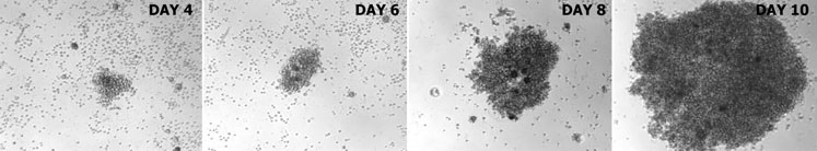
- Leave the plates in the incubator. The colonies will appear between 7 to 10 days
Appendix I
Culture Medium A:
RPMI 1640 medium with L-Glutamine (bicarbonate buffered) (Lonza Ref. BE12-702F)
+ 10% FBS (Genycell Ref. GCS0101-500)
+ Penicillin (100U/ml)/Streptomycin (100mg/l) (Gibco Ref. 15070-063)
Culture Medium A+:
RPMI 1640 medium with L-Glutamine (bicarbonate buffered) (Lonza Ref. BE12-702F)
+10% FBS (Genycell Ref. GCS0101-500)
+ Penicillin (100U/ml)/Streptomycin (100mg/l) (Gibco Ref. 15070-063)
+ Ultroser G (1%) (Pall Ref. 15950-017)
Culture Medium B (no FBS):
RPMI 1640 Medium with L-Glutamine (Lonza Ref. BE12-702F)
+ Penicillin (100U/ml)/Streptomycin (100mg/l) (Gibco Ref. 15070-063)
Culture Medium C:
RPMI 1640 Medium with L-Glutamine (Lonza Ref. BE12-702F)
+ 10% FBS (Genycell Ref. GCS0101-500)
+ Penicillin (100U/ml)/Streptomycin (100mg/l) (Gibco Ref. 15070-063)
Culture Medium D:
RPMI 1640 medium with L-Glutamine (bicarbonate buffered) (Lonza Ref. BE12-702F)
+ 10% FBS (Genycell Ref. GCS0101-500)
+ Penicillin (100U/ml)/Streptomycin (100mg/l) (Gibco Ref. 15070-063)
+ Ultroser G (1%) (Pall Ref. 15950-017)
+ HAT (Hypoxathine, Aminopterin, Thymidine) supplement which is usually 50X (dilute 10ml in 500mls of medium) (Gibco Ref. 21060-017)